It’s important to understand that early brain development is significant as it sets the stage for your child’s future learning, behavior, and even health. The significance of neurosonography in the first trimester is that it helps track those first critical stages of brain formation and development, ensuring that your baby is starting life on the right path.
The Intricacies of Fetal Brain Development
During the first trimester, several key processes are happening. Initially, the neural plate forms. This then folds to become the neural tube, a critical step in developing your baby’s central nervous system. This period is characterized by rapid neuronal growth, meaning nerve cells are forming at an astonishing rate.
Moreover, another remarkable fact about fetal brain development during this time is the brain-to-body weight comparison. The brain is quite big relative to the rest of the body, reflecting its rapid growth and importance. Understanding these early stages helps show why early neurosonographic screening is crucial for monitoring proper development.
Decoding First-Trimester Neurosonography
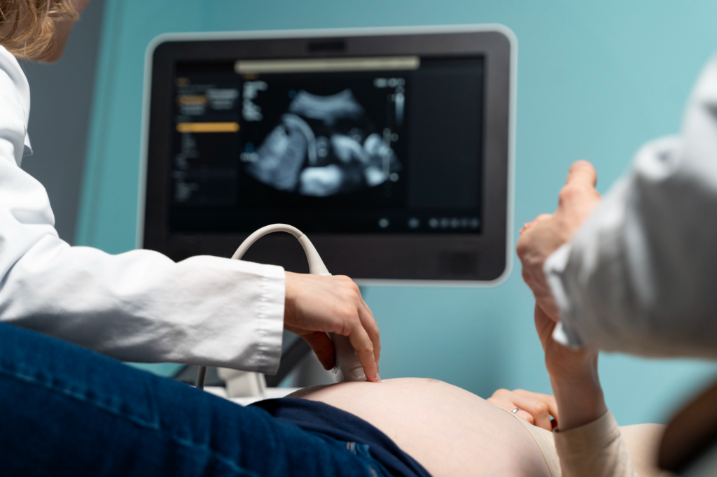
So, what exactly is neurosonography? In simple terms, it’s an ultrasound focused on screening the fetal brain and neurons. There are two common first-trimester neurosonography techniques used: transabdominal and transvaginal.
The transabdominal technique involves placing the probe on the mother’s abdomen, providing a broad overview of the abdominal and fetal structures, while transvaginal ultrasound involves inserting a probe internally, offering more detailed images specific to brain and pelvic structures.
Each method serves different purposes, and medical practitioners choose based on what suits best for monitoring brain health during these crucial weeks.
Timing is Everything: When to Schedule a Neurosonography
Optimal timing is essential when capturing images of your baby’s developing brain. The importance of early neurosonographic screening lies in performing it at the right time for the best results. Between 14 and 16 weeks, neurosonography gives detailed insights.
At these weeks, you may be able to visualize important structures of your baby’s brain, which might not show clearly earlier. While you can get some pictures sooner, the clarity and significance of neurosonography in the first trimester are most effective at this time.
Here is why scheduling your neurosonography is super essential:
- Early insight ensures everything is progressing well.
- Detects potential issues early on.
- Provides peace of mind for expecting parents.
Inside a Neurosonography: Techniques and Evaluations
During the First-Trimester Neuro Sonography, specialists use multiplanar imaging to look at different views of the fetal brain. This approach helps in getting a complete picture of the brain’s progress.
Experts focus on key fetal brain structures such as the hemisphere and the middle line structure. Evaluating these elements and correlating them with the baby’s gestational age is crucial in providing accurate insights into brain health.
Understanding what’s going on during these scans goes a long way in showing why first-trimester neurosonography techniques and their evaluations are vital for both parents and healthcare providers.
Significant Findings: Common Fetal Brain Anomalies
Though the experience is often reassuring, neurosonography can sometimes uncover anomalies. Common abnormalities identifiable include acrania, where the skull is partially missing, and holoprosencephaly, which affects the brain’s structure.
Other potential issues detected include neural tube defects, which might need further investigations. As daunting as it sounds, many findings suggest manageable conditions. Knowing this provides reassurance, paving the way for the right medical approaches and support.
Complementing Diagnostics: The Role of Fetal MRI
While First-Trimester Neuro Sonography offers lots of insights, sometimes a fetal MRI is suggested as a supplementary tool. This is because MRIs may provide additional details when looking deeper into any detected issues. The expertise required for analyzing these imaging processes further solidifies their importance in comprehensive prenatal care.
External Influences: Maternal and Environmental Factors
It’s not just all about what science can do; mothers have an influential role. Lifestyle choices significantly affect fetal brain health. Good nutrition, avoiding infections, and maintaining healthy habits can positively shape prenatal development.
Here are simple steps for supporting baby’s growth:
- Eat a balanced diet.
- Follow regular health check-ups.
- Avoiding harmful substance use.
By understanding these factors, parents can support brain development through simple everyday choices.
Establishing Guidelines: Expert Recommendations
Clear guidelines set by professionals, such as those from ISUOG, provide recommendations on first-trimester neurosonography. They offer criteria for when detailed screenings are advisable.
While there are minor risks in early monitoring, the benefits tend to outweigh them. Parents gain crucial insights into the baby’s development and prepare to tackle potential challenges with foresight.
Wrapping Up: Final Thoughts for Expecting Parents
By now, you’ve likely gained an understanding of the importance of early neurosonographic screening. Keep in mind that proactive monitoring is beneficial for your baby’s healthy start.
Embrace these insights, stay informed, and don’t hesitate to speak with healthcare providers about any concerns or procedures.
Best wishes for a joyful and healthy journey ahead! Your proactive steps today pave the way for a brighter future for your little one.